
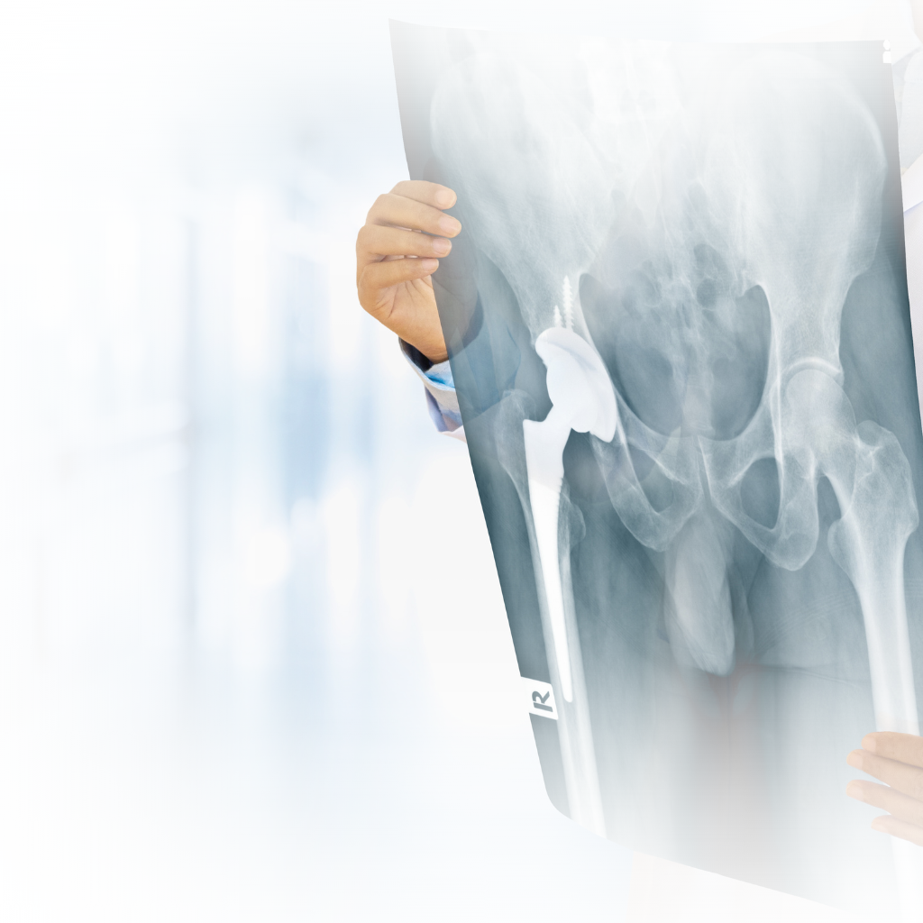
Orthopedic X-rays, also known as orthopedic radiographs, are imaging tests commonly used in orthopedics to visualize bones, joints, and related structures. X-rays provide detailed images that help diagnose various orthopedic conditions, including fractures, dislocations, arthritis, bone tumors, and degenerative changes. We have on-site digital imaging and strong relationships with other specialty care providers to provide diagnostic tools and treatment plans for those of all ages.
Preparation: There is usually no specific preparation required for an orthopedic X-ray. You may be asked to remove any jewelry, metal objects, or clothing that could interfere with the X-ray images.
Positioning: You will be positioned on an X-ray table or stand, and the technologist will guide you into the appropriate position to obtain the desired views. Depending on the area being examined, you may be asked to stand, sit, lie down, or move into different positions.
Protective Measures: In some cases, a lead apron or shield may be placed over parts of your body that are not being imaged to minimize radiation exposure.
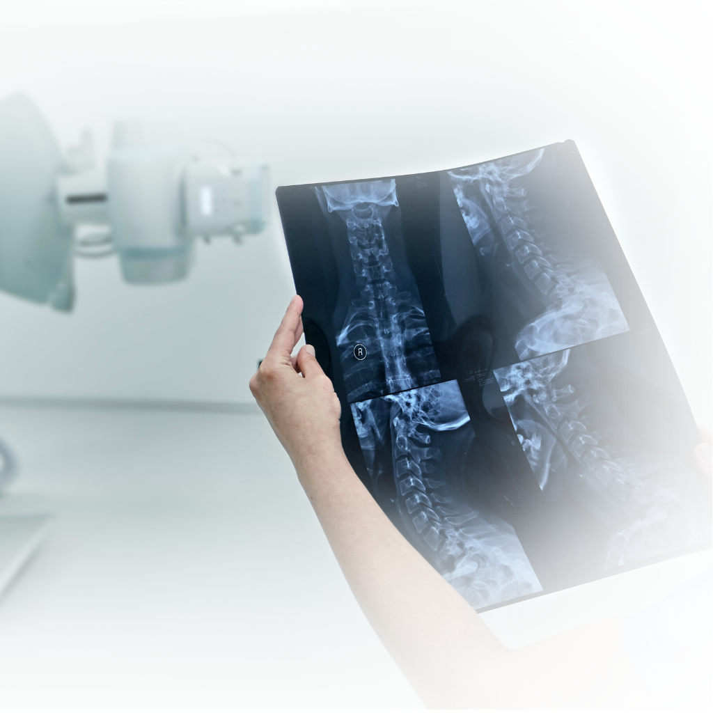
X-ray Machine: The X-ray machine, operated by a radiologic technologist, will be positioned to generate the X-ray beams. You will be instructed to remain still and hold your position while the images are taken.
Multiple Views: Depending on the area being examined, multiple X-ray views may be necessary to capture different angles and perspectives. This helps provide a comprehensive evaluation of the bone or joint.
Image Review: After the X-rays are taken, the images will be reviewed by a radiologist, who is a specialized physician trained to interpret medical images. The radiologist will analyze the X-rays and provide a report to your orthopedic doctor.
Orthopedic X-rays offer several benefits, including:
Quick and non-invasive: X-rays are typically quick to perform, and the results are available shortly after the imaging procedure.
High-quality images: X-rays provide detailed images of bones, joints, and fractures, allowing for accurate diagnosis and treatment planning.
Cost-effective: X-rays are generally more affordable compared to other imaging techniques.
However, it’s important to note that X-rays have some limitations:
Limited soft tissue visualization: X-rays primarily show bones and are less effective in visualizing soft tissues, such as muscles, tendons, and ligaments.
Radiation exposure: X-rays involve exposure to ionizing radiation. While the amount of radiation is relatively low, pregnant women and children are more sensitive to radiation and precautions may be taken to minimize their exposure.
It’s important to consult with an orthopedic specialist who can determine whether an X-ray is necessary for your specific condition. They will assess your symptoms, perform a physical examination, and consider other factors before recommending any imaging tests. The orthopedic doctor will use the X-ray results, along with other clinical information, to provide an accurate diagnosis and develop an appropriate treatment plan.
Pelham, AL
info@albjc.com
205.621.4835
205.621.3778