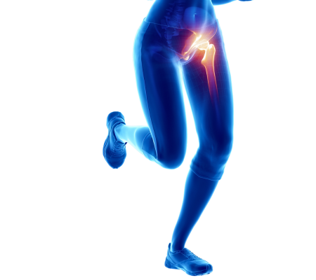
Hip arthroscopy is a minimally invasive surgical procedure used to diagnose and treat various hip joint conditions. It involves the insertion of a small camera, called an arthroscope, into the hip joint through small incisions. The arthroscope allows the surgeon to visualize the inside of the hip joint and perform necessary surgical interventions.
Here is an overview of the hip arthroscopy procedure:
Preoperative Assessment:
Before the surgery, the patient undergoes a comprehensive evaluation, including a medical history review, physical examination, and imaging tests such as X-rays, MRI, or CT scans. These assessments help determine the appropriateness of hip arthroscopy and plan the procedure accordingly.
Anesthesia:
Hip arthroscopy is typically performed under general anesthesia, which means the patient is unconscious during the surgery. In some cases, regional anesthesia or a combination of general and regional anesthesia may be used.
Incisions:
Several small incisions, typically around 1 cm in length, are made around the hip joint. These incisions serve as entry points for the arthroscope and other surgical instruments.
Insertion of the Arthroscope:
The arthroscope, a thin tube with a camera attached to the end, is inserted into the hip joint through one of the incisions. The camera allows the surgeon to visualize the joint structures on a monitor.
Evaluation and Diagnosis:
The surgeon carefully examines the inside of the hip joint, including the articular surfaces, labrum, cartilage, ligaments, and tendons. Any abnormalities, such as labral tears, cartilage damage, impingements, loose bodies, or synovitis, can be identified and assessed.
Surgical Interventions:
Once the problem areas are identified, the surgeon can perform various surgical interventions using specialized instruments inserted through the additional incisions. These interventions may include:
Labral Repair or Debridement:
The labrum, a ring of cartilage around the rim of the hip socket, may be repaired or trimmed to address tears or fraying.
Cartilage Repair or Microfracture:
If there are areas of cartilage damage, the surgeon may perform techniques to stimulate the growth of new cartilage or remove loose cartilage fragments.
Impingement Correction:
If there is evidence of femoroacetabular impingement (FAI), the surgeon can shave or reshape the bone to improve joint clearance.
Removal of Loose Bodies:
If there are loose fragments of bone or cartilage within the joint, they can be removed during hip arthroscopy.
Closure:
Once the necessary interventions are performed, the surgeon closes the incisions using sutures or adhesive strips.
Postoperative Care and Rehabilitation:
After the procedure, the patient is monitored in a recovery area before being transferred to a hospital room. Pain management, early mobilization, and physical therapy are crucial for recovery and rehabilitation. Physical therapy helps restore strength, range of motion, and overall function.
The recovery period after hip arthroscopy is generally faster compared to traditional open surgery, but it still requires a period of rehabilitation and gradual return to activities. The specific rehabilitation plan will depend on the individual’s condition and the procedures performed during arthroscopy.
It’s important to note that not all hip conditions can be effectively treated with hip arthroscopy. The suitability of this procedure depends on the specific diagnosis and patient factors. An orthopedic surgeon specializing in hip arthroscopy can evaluate your specific case and determine if hip arthroscopy is the most appropriate treatment option for you.
As with any surgical procedure, hip arthroscopy carries certain risks and potential complications, including infection, bleeding, nerve injury, blood clots, or ongoing hip pain. Your surgeon will discuss these risks when you are seen by a specialists at Alabama Bone and Joint Clinic. Call today to schedule an appointment with one of our experts in orthopedic care.